4D Sonography
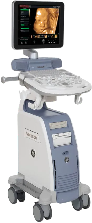
Machine
GE Voluson P8
We use best-in-class US FDA approved 4D ultrasound and color doppler machine.
Sonography is a boon for pregnant women. Sound waves are used to investigate the fetus. It is completely safe technology. Sonography poses no risk to the fetus or the mother.
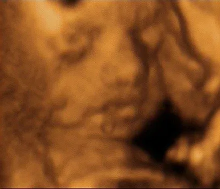
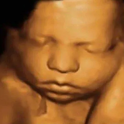
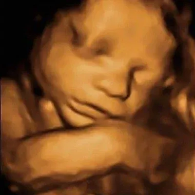
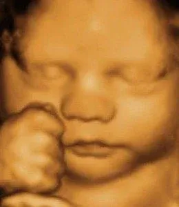
Using our 4D machine we are able to create life-like images of the baby.
4D Sonography Images
When and why should sonography be done during pregnancy?
1) First 7 to 10 weeks
- Is the implantation proper?
- Is the fetal growth regular?
- How many fetuses (twins, etc.)?
- Is there bleeding around the fetus?
- Is there an ectopic pregnancy?
2) 11 to 13 weeks
Is there any possibility of a genetic defect, e.g. Down's Syndrome? Nuchal translucency and nasal bone are examined.
3) 18 to 20 weeks
All organs of the fetus — brain, spine, chest, abdominal organs, kidneys, hands and feet — are inspected for any defects.
4) 30 to 42 weeks — to see the growth of the baby
Baby's weight, amount of amniotic fluid, baby's movement, and baby's blood supply are inspected. Recommended if there were complications in a previous pregnancy, if amniotic fluid decreases, or if there is diabetes.
Sonography Schedule
| Weeks | Purpose |
|---|---|
| 7 – 10 | Confirm implantation, fetal count, rule out ectopic pregnancy |
| 11 – 13 * | Nuchal translucency — Down's Syndrome screening |
| 18 – 20 * | Anomaly scan — all fetal organs |
| 30 – 42 | Growth scan — weight, amniotic fluid, movement, blood supply |
* most important